An Insight Into The Structure Of Ovarian Follicles
June 24, 2018The cortex of a mature ovary contains numerous spherical or oval sac like masses of cells like the island of cells called ovarian follicles, at various stages of maturation. During the embryonic development stage, millions of germ other cells or oogonia are formed within each fetal ovary. However no more oogonia are formed and added after the birth of the girl child. The oogonia divides by mitosis to form a primary oocyte.
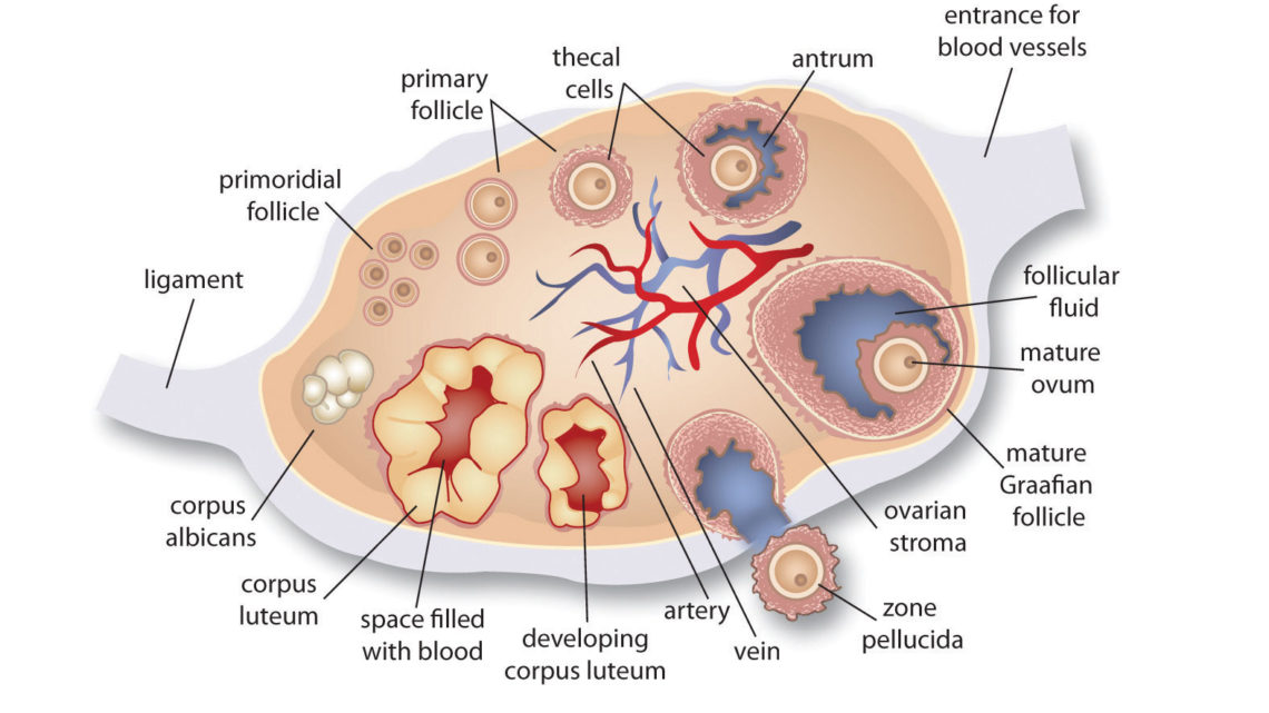
Each primary oocyte gets surrounded by a layer of granulosa cells to form primordial follicle. Let us look into the
- Primordial follicles: These are immature follicles each primordial follicle has a primary oocyte arrested at a particular stage of meiotic cell division. And it is surrounded by a single layer of granulosa cells or follicle cells.
- Primary follicle: These are maturing follicles, and each has a primary oocyte. The primordial follicle first develops into a primary follicle and then by the division of granulosa cells it forms multiple layers of granulosa cells around it.
- Secondary follicle: After the completion of meiotic cell division the secondary oocyte is formed which is surrounded by multiple layers of granulosa cells. The granulosa cells start secreting estrogen and a cavity or antrum is filled with fluid which forms within the follicle. The entire follicle is then surrounded by a connective tissue. The tissue covering is known as the theca, and it derives itself from the stroma. A lot of pregnancy magazines in Hindi give a detailed account of the different kinds of follicles which are present in the ovaries of women, and how they mature at each stage to make a pregnancy successful.
- Mature follicle: This is a fully grown Graafian follicle with double layered theca, surrounding a double layered granulosa cell, separated by continuous fluid filled with antrum. Secondary oocyte is arrested at a particular stage of meiotic cell division and it lies within the inner layer of granulosa cells. From this follicle the secondary oocyte is shed by ovulation which occurs nearly fourteen days before the onset of the next menstrual cycle.
- Corpus luteum: After the extrusion of the ovum whatever remains of the Graafian follicle is called the corpus luteum. It is a yellow colored follicle filled with granulosa cells containing yellow lutein pigments.
- Pregnancy magazine in Hindi let the pregnant mothers know, what happens at the end of the functional life of the corpus luteum. It completely degenerates and becomes converted into a mass of fibrous tissue without the lutein pigments, and is known as the corpus albicans or the white body.
Functions of the ovary:
Ovary is a mixed gland and it performs both exocrine and endocrine function. The exocrine function of ovary is the production and maturation of female gametes or ova. The endocrine function of ovary is secretion of hormones. The granulosa cells of the maturing follicles of ovary secretes estrogen hormone and the corpus luteum secretes both, estrogen and progesterone hormone, for the maintenance of pregnancy. The granulosa cells are divided into two layers, due to the presence of a fluid filled cavity called antrum.


